CALL or BOOK an APPOINTMENT TODAY !!!!
703-276-1010 Located 2 blocks from Ballston METRO STATION
Open MON - FRI From 8:00 am to 5:00 pm
CALL or BOOK an APPOINTMENT TODAY !!!!
Open MON - FRI From 8:00 am to 4:00 pm
Free Gingival Graft
Free Gingival Graft
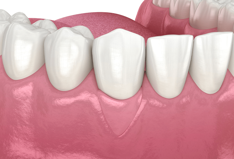
Gingival graft refers to a keratinized epithelial-connective tissue detached from its original site to be grafted in an area other than the donor area.The gum graft is a microsurgery that is used to recover damaged gums. However, tissue loss gradually exposes the tooth, which can end up affecting the bone that supports the teeth.It is performed by taking the patient’s soft tissue and placing it in the area of the tooth or implant that has been uncovered by gum recession.
This graft consists of a fragment of connective epithelium obtained from the palate.It is not a predictable coverage technique for complete root coverage because graft survival on the avascular root surface depends exclusively on the vascular bridges formed between the grafted tissue and the remaining periosteal bed around the root exposure.Therefore, to increase the survival success of the graft on the root, it is necessary to cover at least 3 mm of the mesial, distal and apical periosteal bed of the bone dehiscence. As a result, the gingival graft can only be used to cover shallow and shallow recessions.
What is the free gingival graft?
Gingival grafting represents the latest choice among root coverage techniques. It is most often used to treat small teeth in the lower arch, especially incisors, and when the gums around the neighboring teeth are light pink. The gingival graft is an effective technique when the braces inserted on the gingival margin determine the mobilization of the marginal tissue. The goal is to make the keratinized tissue along the edge taller and thicker. In this way, it facilitates the control of oral hygiene by the patient, thus reducing the accumulation of subgingival bacterial plaque.
Indications for free gingival grafting
The gingival graft is indicated when desired to increase keratinized tissue in height and especially in thickness in the presence of small root exposures.This last requirement is verified, for example, in patients who must undergo orthodontic
treatments where dental vestibularizations are going to be performed.The main objective is to provide a dense gingival margin made up of keratinized tissue to the receded tooth.
Free gingival graft in implantology
Free gingival grafting associated with implantology has been recommended for areas without keratinized gingiva around dental implants.

1. Cover Expose Root of teeth
Receding gums expose the roots, promoting tooth decay and other oral health issues. In severe circumstances, this might lead to tooth loss. A gum transplant protects teeth and roots from decay and inflammation, eliminating receding gums.

2. Reduces Tooth Sensitivity
When the tooth's roots are exposed, they develop extreme sensitivity to heat and cold. Gum grafting surgery can provide more protection, lessen annoying pain, and eliminate the dental sensitivity that comes from gum recession.

3. Enhance your smile beauty
Your teeth may appear longer and less beautiful if your gums are receding. Your smile's cosmetics can be greatly enhanced by adding new gum tissue by grafting since your gums will appear more even, natural, and in general more attractive.

4. Recovery
Plaque can get into the gums and other soft areas if the gum tissue isn't strong and healthy. This can lead to decay and cavities. As a result, there is bone loss, and subsequent gum tissue deterioration. Gum grafting strengthens the gum tissue and enhances periodontal health.
Facing Gum Recession? Gingival Grafting is an ideal option.
You might wish to discuss your treatment options with a periodontist if you have thin, receding gums. After all, gum recession that is left untreated increases the risk of getting gum disease or tooth decay. Additionally, a smile that is uneven might be caused by gums that have pulled away from your teeth.A free gingival graft is one option that you might be asked to consider. Our highly skilled periodontists at Virginia Dental Care execute a free gingival graft to return your gums to a healthier state permanently.
Why is it important to have keratinized gums?
A good keratinized gum retains plaque bacteria and improves the gingival health of the
surrounding tooth or implant.
The free gingival graft will have the following main goals:
First, increase the width of the attached gingiva around teeth and dental implants.
And covering the roots exposed by gingival recession, eliminating the insertion of potentially
harmful braces.
Surgical technique
How Long Does a Free Gingival Graft Take to Heal?
Gum graft healing
The healing of the gingival graft is characterized by an initial phase of hospitalization that is clinically manifested by the appearance of a white layer on the external surface of the graft. After a few days, approximately five or seven, the whitish layer begins to disappear, revealing a reddish tissue with little consistency. In this phase, it is very important not to traumatize the tissue, so it is advisable not to use a toothbrush for hygiene, continuing with the chemical control of bacterial plaque using chlorhexidine rinses. Once the epithelialization phase is over (after 14 days), the re-epithelialization of the connective tissue begins from the surrounding epithelial tissue. Re-epithelialization manifests clinically with the appearance of consistent tissue characterized by a pink surface that will lighten in color over time. It should be remembered that the characteristics of keratinization of tissue depend on the properties of the subepithelial connective tissue. Therefore, the gingival graft will acquire chromatic and superficial characteristics equal to those of the area of the palatal fibro mucosa where it was obtained.
Gingival Graft Considerations
Gingival grafting represents the latest choice among root coverage techniques. It’s hard to predict when all of the roots will cover the soil, especially in wide, deep recessions. In addition, the aesthetic result is unsatisfactory due to the difference in the color of the grafted tissue to the adjacent soft tissues and the unevenness of the mucogingival junction. It may be used to treat small recessions in the lower dental arch, especially in the front teeth, and when the gums of the teeth next to the recession are a light pink color. The gingival graft is an effective technique when the braces inserted on the gingival margin determine the mobilization of the marginal tissue. The goal is to increase the height and thickness of the marginal keratinized tissue.
Gingival graft contraindications
This type of graft is contraindicated in the following cases:
1. Patients with high aesthetic demand
2. When the recessions are wide and deep
3. When there is deep vestibular probing associated with gingival recessions

What are the objectives of the gum graft on implants?
Soft tissue manipulation seeks good aesthetics and better maintenance of the health of dental implants.
Complications involving the soft tissue surrounding dental implants, such as mucosal irritation, gingival hyperplasia, peri-implantitis, and insufficient epithelial adhesion, are conditions that can alter the prognosis and longevity of Osseointegrated implants.
Establishing aesthetics and harmony around restoration is a great challenge.
The gum that surrounds the implant in its structural form is the one that most resembles the gum that surrounds the natural tooth.
The main difference between this tissue about the tooth and the implant resides in the way of interfacing with the gingival tissue and the surrounding bone, that is, the absence of cement around the dental implants.
Why is the quality of the gum around your implants important?
The importance of the quality of the gums around the implants does not come from a purely aesthetic need. There is a relationship between keratinized gingiva around implants and their long-term success. In other words, when you have good keratinized gingiva around your implants, there is less pocket depth and a better prognosis. Different methods, such as free gingival grafting, connective tissue grafting, acellular matrix, and
apical repositioning of the buccal flap, have been used to make keratinized peri-implant tissue, which is easier to care for and more resistant to mechanical stress. An increase in keratinized gingiva at the time of reopening with the apical repositioning technique of the divided vestibular flap could achieve several objectives with a single procedure:
1. Visualization of the dental implant.
2. The increased strip of keratinized gum.
3. Deepening of the vestibule.
4. Improved aesthetics with increased vestibular soft tissue thickness.
The most widely used technique is that of subepithelial tissue graft removed from the palate or the maxillary tuberosity. It treats moderate horizontal or vertical defects, such that connective tissue is placed under a split labial flap. The presence of a resistant mucosa favors a clinical situation in which it is easier to keep the tissues free of bacterial plaque. And with a good keratinized gum, the peri-implant soft tissues’ health remains free of inflammation for a long time.


